Advanced Cytogenetic Technique – Microarray Testing

Advanced Cytogenetic Technique: Microarray Testing

• 20% of stillborn babies have a major malformation
• 3% of live-births have congenital anomalies
• Diagnosis of a malformation requires:
– A number of “broad spectrum” analysis
– Followed by more sophisticated, accurate and targeted tests
• Detailed ultrasound and invasive fetal testing
– Etiology
– Prognosis
– Recurrence risk
– Prevention / options in future pregnancies


Genomic microarray technology
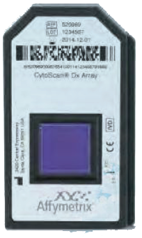
Each microarray contains 2.6 million markers for copy
number & 750,000 SNPs markers
Yield is 15-20%
Whole genome- High resolution

ACMG Recommends Replacing Karyotyping with Chromosomal Microarrays as
‘First-Line’ Postnatal Test
“Increased resolution of microarray technology over conventional cytogenetic
analysis allows for identification of chromosomal imbalances with greater
precision, accuracy and technical sensitivity.”


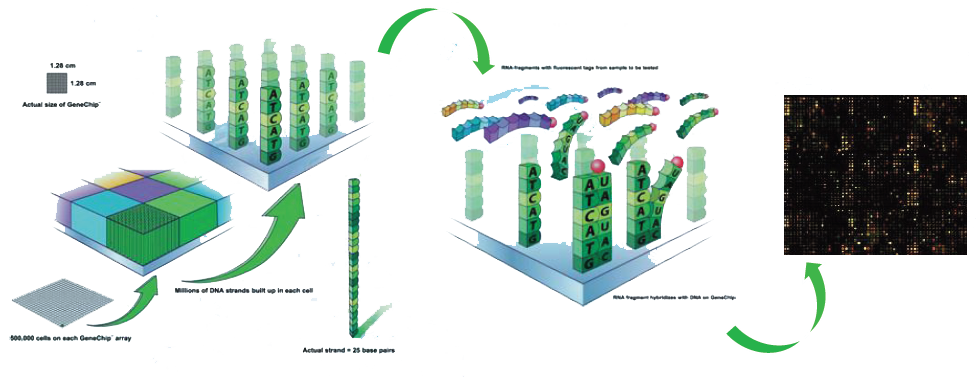
Affymetrix-25 mers are in-situ synthesized on a glass
wafer nucleotide by nucleotide using photolithography


• Advanced maternal age
• Previous child with de novo chromosome aneuploidy
– Woman 30 years, child with T21: Increased Recurrence risk for any
chromosome abnormality (1/100) versus age-related risk (1/390)
• Parental structural chromosome abnormality
• Family history of genetic disorder
• Elevated risk based on maternal screening
• Foetuses with abnormal ultrasound findings

Based on the increased detection of clinically relevant abnormalities in both structurally normal and abnormal pregnancies, Chromosomal Microarray Analysis (CMA) should be transitioned to become the First Tier Test for invasive prenatal diagnosis.
Source: New England Journal of medicine,Vol 367,No 23,Dec.2012

• CMA detects chromosome abnormalities and new genetic syndromes which would be missed by conventional cytogenetics
• Most Copy Number Variations can be interpreted based on gene content, size, inheritance, databases
• Counseling issues are not unique to prenatal CMA
• As CMA transitions into clinical practice counseling by professional with knowledge and expertise in CMA will be required
• Need large databases of array findings and associated phenotype from “unbiased populations”

• If a fetal structural anomaly is identified on ultrasound examination, invasive prenatal diagnosis should be offered
• A negative cell free fetal DNA test result does not ensure an unaffected pregnancy
• A patient with a positive test result should be referred for genetic counseling and offered invasive prenatal diagnosis for confirmation of test results
• Cell free fetal DNA does not replace the accuracy and diagnostic precision of prenatal diagnosis with CVS or amniocentesis, which remain an option for women

1. In patients with a fetus with one or more major structural abnormalities identified by ultrasound who are undergoing invasive prenatal diagnosis, Chromosomal Microarray Analysis is
recommended. This replaces traditional fetal karyotype – which may be viewed as a low-resolution whole genome analysis.
2. In patients with a structurally normal fetus undergoing invasive prenatal diagnostic testing, either traditional chromosome analysis or chromosome microarray analysis may be performed.
3. As most copy number mutations identified by Chromosomal Microarray Analysis are not associated with increasing maternal age, the use of CMA for prenatal diagnosis should not be
restricted to women aged 35 and older.
4. In case of intrauterine fetal demise or stillbirth, when further cytogenetic analysis is desired, Chromosomal Microarray Analysis on fetal tissue is recommended. This increases the likelihood
of obtaining results and improves the detection of causative abnormalities.

• Approximately 60-70% of first trimester miscarriages are being caused by chromosomal abnormalities
• Traditional cytogenetic analysis of these samples is challenging due to high rates of culture failure and maternal contamination
• Chromosomal microarray analysis overcomes these limitations and has proven to be an excellent tool for detection of chromosomal aberrations in these samples
Ref. : Wang et al.Molecular cytogenetics 2014 7:33


