RENAL PATHOLOGY AND ELECTRON MICROSCOPY
Department of Renal Pathology & Electron Microscopy
• Only stand alone department of its type in India handling one of the largest renal biopsy workload in the world.
• Among the very few centres in Asia to offer all the three diagnostic modalities for renal biopsies (light microscopy, direct immunofluorescence (DIF) and Electron Microscopy) under one roof.
• First & only diagnostic biological transmission electron microscopy facility in India to be accredited by National Accreditation Board for Testing and Calibration Laboratories (NABL).
• Providing & maintaining highest standards of quality and promptness of service are the key features of our practice.
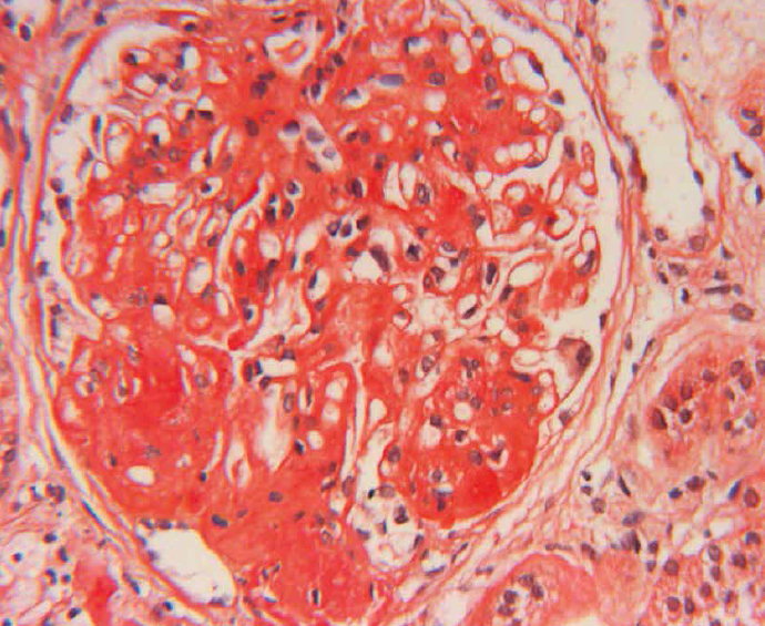
LIGHT MICROSCOPY & IMMUNOHISTO-CHEMISTRY
• Each renal biopsy is examined at multiple serial levels and supplemented with a battery of routine & special stains.
• A panel of ancillary techniques & markers is employed upfront for optimal diagnosis at no additional cost.
• We provide referral services and facility of specialized tests for wide spectrum of renal diseases
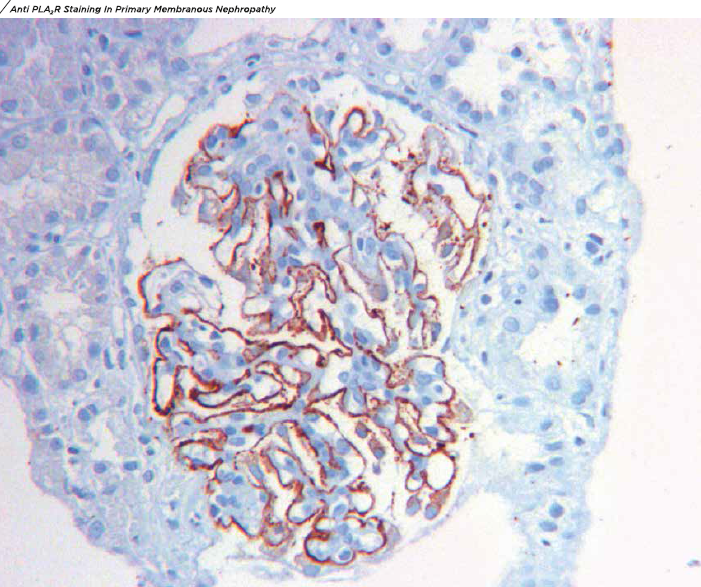
1. Phospholipase A2 receptor (PLA, R) and Thrombospondin type 1 domain containing 7A (THSD7A)
Recently characterized markers of Primary Membranous Nephropathy (MGN). Staining for PLA, R and THSD7A is done routinely for all cases of Membranous glomerulopathy, helping the Nephrologists in deciding optimal treatment strategies. [show_more more=”Continue Reading ▼” less=”Show Less ▲”]
2. Typing of amyloid on renal biopsies
• Primary (AL) amyloidosis by Kappa & Lambda light chain.
• Amyloid associated protein (SAA) for secondary (AA) amyloidosis.
• Leukocyte Chemoattractant Protein 2 (LECT2).
3. DNA J Heat Shock Protein Family (Hsp40) Member B9 (DNAJB9) is a recently described marker for Fibrillary glomerulonephrItis. We are the first and only centre in India to offer staining for DNAJB9 in renal biopsies.
4. Type Ill collagen – A gold standard confirmatory test for diagnosis of Collagenofibrotic Glomerulopathy.
5. Fibronectin – Confirmatory test for this unusual glomerular disease (Flbronectln Glomerulopathy).
6. IgG4 – For diagnosis of IgG4 associated Renal disease, characterized by IgG4 positive plasma cell rich tubulointerstitial nephritis.
RENAL TRANSPLANT PATHOLOGY
• Formidable reputation as a reference centre for renal allograft biopsy interpretation.
• Verbal reports of renal allograft biopsies are communicated personally to the referring physician by our Renal Pathologists within 24 hours.
• All the necessary ancillary markers required for a reasonable opinion are performed upfront and at no additional cost.
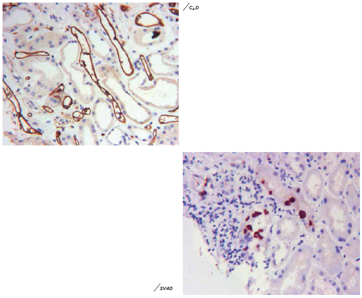
/ C4D, SV4O (BK virus) sta n ng performed for all the transplant biopsies.
/CYTOMEGALOVIRUS (CMV), EPSTEIN BARR VIRUS ( EBV), CD3, CD2O etc. applied as per the need of the case.
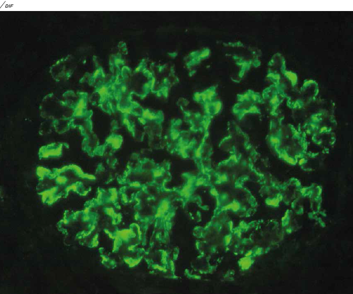
DIRECT IMMUNOFLUOROSCENE
• Comprehensive panel for Direct immunofibrinogen (DIF) studies including immunoglobulins (IgA, IgG, IgM), early and late complement components (C3, Clq) Fibrinogen and light chains (kappa & lambda) supplemented by several other markers for all the cases.
• Subtyping of IgG classes (IgG1, tgG2, tgG3 & IgG4) is performed wherever it is required.
• DIF studies on Pronase/Proteinase treated sections obtained from paraffin-embedded tissue :
Useful in cases where DIF tissue is either inadequate or not available and also helps in cases where unmasking of specific antigens is part of the diagnostic algorithm.
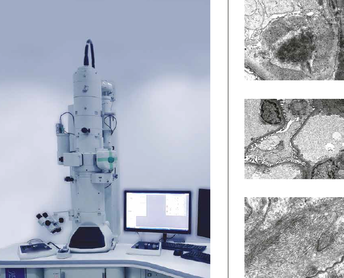
ELECTRON MICROSCOPY
• Electron Microscopy is an indispensable technique required for optimal analysis of renal biopsies.
• Dr Lal Pathlabs the first private standalone laboratory in India to setup an in house Transmission Electron Microscopy (TEM) facility.
• Our Laboratory has capability to perform Transmission Electron Microscopy on tissue retrieved from paraffin block prepared for routine his to pathology studies. This technique enables ultrastructural analysis in cases where fresh tissue is either not available or cannot be procured due to clinical & technical limitations.
• TEM facility houses a dedicated state of the art electron microscopy sample processing laboratory and the JEOL 120 kv Transmission Electron Microscope.
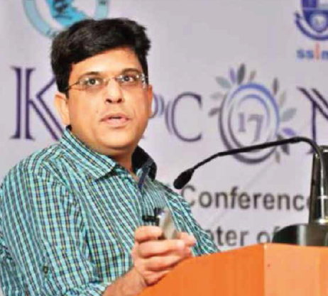
Dr. Alok Sharma
• Dr. Alok Sharma is among the most respected & accomplished Renal Pathologists recognized in India and spearheads the team of Renal Pathology & Electron Microscopy department at the National Reference Laboratory (NRL) of Dr. Lal Pathlabs, New Delhi.
• Trained in Renal Pathology at the All India Institute of Medical Sciences (AIIMS), New Delhi and a Visiting Clinician in Renal Pathology & Electron Microscopy at the Mayo Clinic, USA.
• Credited with establishing one of the largest Renal Pathology departments in the world at Dr Lal Pathlabs Ltd, New Delhi and reporting more than 35000 renal biopsies in his illustrious career.
• Authored more than 100 research publications in various peer-reviewed national & international journals including some landmark papers in the field of Renal Transplantation Pathology.
• Recipient of many prestigious awards including the HD Tandon Memorial award, Dr. V Ramalingaswamy Memorial award and the Dr. VR Khanolkar National award for the best-published research work in the field of Pathology.
• Creator and webmaster of the globally acclaimed and among the most widely accessed websites on Renal Pathology in the world www.nephro-pathology.com
MAJOR TEST PANELS OFFERED
| S No | Test Panel Name | Code | TAT | Comments |
| 1 | Kidney biopsy panel 1 (includes light microscopy with special stains + Direct Immunofluoroscence) | Z 353 | 5 working days | Verbal impression communicated earlier in urgent cases |
| 2 | Kidney biopsy panel 2 (includes light microscopy with special stains) | Z 355 | 5 working days | Verbal impression communicated earlier in urgent cases |
| 3 | Kidney biopsy panel 3 (includes light microscopy with special stains + Direct Immunolluoroscence + Transmission Electron Microscopy) | Z 830 | 10 working days | Verbal impression communicated earlier in urgent cases & L M & DIP report released earlier wherever necessary |
| 4 | Electron Microscopy | J 154 | 10 working days | TEM can also be performed on paraffin embedded tissue |
| 5 | Kidney biopsy Direct immunofluor- oscence panel (Panel of IgA, IgG, IgM, C3, Clq, kappa light chains and lambda light chains | Z 829 | 3 working days | Additional markers can be put on request within 3 days |
[/show_more]