What are the different types of X-rays and their usage?

X-ray is a shorthand for X-radiation. Discovered and named by Wilhelm Rontgen, x-rays can neither be see with the naked eyes nor can they be felt. The main aim behind the discovery of this technology was to study the internal parts of the human body, which cannot be seen otherwise. The human system is a complex structure of mass that comprises of a plethora of organs and bones which perform a colossal range of day-to-day activities. Due to some internal or external factors, the system faces issues. This is where an X-ray test helps in detecting problems by producing images. From dental to chest, abdomen to ankle every part of the body can be studied with the help of X-ray.
Types of X-rays
Medical science recognizes different types of X-rays. These are as follows:-
Standard Computed Tomography
A standard computed tomography or otherwise known as computerized axial tomography is performed in a hospital or at a radiologist’s office. The test aids in obtaining detailed images of areas inside the body, typically for the diagnosis of circulatory system such as blood vessel aneurysms, blood clots, and coronary artery disease.
Kidney, Ureter, and Bladder X-ray
Also known as KUB X-Rays, it is performed to assess the abdominal area for causes of abdominal pain, or to assess the organs and structures of the urinary and/or gastrointestinal (GI) system. A KUB X-ray is practically the first diagnostic procedure used for the assessment of the urinary system.
Teeth and bones X-rays
These x-rays give a high level of detail about bones, teeth, and supporting tissues of the mouth. These X-rays enable dentists to look at the tooth roots, the status of the developing tooth, and health of the bony areas.
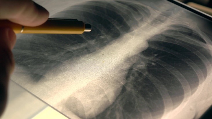
Chest X-rays
A small test that uses radiation to produce images of the bones, tissues, and organs of the body. The doctor prescribes a chest X-ray for a number of reasons like shortness of breath, fever, chest pain, and persistent cough. It is a quick and effective test that aids in analyzing the health of some of the most vital organs.
Lungs X-rays
This type of X-Ray is done to assess the lungs by comparing the upper, middle & lower zones of the lungs. The asymmetry of lung density is represented as either abnormal whiteness (increased density) or abnormal blackness (decreased density). Once you spot the asymmetry, the next step is to decide which side is abnormal. If there is an area that is different from the surrounding ipsilateral lung, then this is likely to be the abnormal area.
Abdomen X-rays
An imaging test to look at structures and organs in the belly, this x-ray encompasses the small and large intestines, liver, and stomach. It is one of the first tests to find a cause of nausea, stomach pain, vomiting, and swelling. Other tests like intravenous pyelography, ultrasound, and CT scan are also done to find more specific problems.
Why is it done?
The X-Ray tests are done to check for broken bones and abnormal activities in the body. It is mostly a quick & painless procedure and a very effective way of looking at the bones and other body parts. Your doctor may prescribe you an X-Ray if you experience prolonged explainable pain in your body; as an X-ray produces images of the affected organs, bones, and tissues of the body. Also, a chest X-Ray may be used to detect pneumonia.
Here are a variety of problems which can be detected with the help of an X-Ray:
- Problems in the chest
- Some kind of lung condition such as chronic obstructive pulmonary disease, cystic fibrosis, lung cancer, pneumonia, and pneumothorax
- Heart Conditions, such as Heart Failure, etc. and for viewing its size & shape
- To monitor progress of the chest area after a surgery
- To find objects like coins or small pieces of metal in the lungs
- Bone Fractures
- Tooth Complications, such as Loose Teeth and Dental Abscesses
- Scoliosis (Irregular Curving of the Spine)
- Cancerous & Non-Cancerous Bone Tumors
- Dysphagia (Difficulty or Discomfort in Swallowing)
- Breast Cancer
How to prepare?
Different types of X-rays speak of different set of procedures and come with their own set of rules and regulations as well. For instance, you need to fast for about 12 hours for getting an exam done in the case of most X-rays. In the case of a dental X-ray, no such preparations are needed. However, there are some rules that remain the same – remove all body piercings, jewellery and other metals before the X-ray. In some special cases, you may also be asked to discontinue some medications, such as during pregnancy or other medical conditions, as radiation can serve harmful to the body.
How is the test taken?
The test is taken by a radiology technologist one from the back and the other one from the side of the body. In an emergency, one image is taken from the front. During the exam, you have to hold your breath so that your chest stays completely still. When the test is completed, you have to wait until the radiologist confirms all the pictures have been obtained. Some hospitals have portable x-rays equipment. If a test is done with a portable x-ray machine, you need to be placed in the correct position by the nurse.
Risks
If you’re worried about the effect of radiation exposure, then most physicians state that X-rays are safe. The kind of radiations sent to the body are safe of the internal system as well. In some cases, a special substance is given in addition to throwing radiation waves, as the combination helps in viewing structures which a normal scan may not be able to reveal. At the same time, it is important to note, and as stated in the paragraph above, X-ray test is not recommended to pregnant women. This is because the rays of the radiation process are harmful for the developing baby. He/she, in the event of strong radiation may develop lifetime abnormalities.
To conclude, different types of X-rays are used for detecting, diagnosing, and analyzing different body conditions. Each of these tests, as listed above, has its own role to play. They are highly effective and quite safe as well.


